
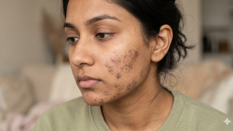
When a client comes in frustrated that their acne is clearing but their skin still doesn’t look right, the answer almost always lies in two conditions that are frequently confused for one another: post-inflammatory hyperpigmentation (PIH) and post-inflammatory erythema (PIE). Both appear after skin inflammation, both leave marks in the wake of a breakout, and both tend to be lumped together under the catch-all of “acne scarring.” But they are fundamentally different in origin, behavior, and critically, in how they should be treated.
For estheticians committed to achieving real, measurable results for their pigmentation clients, the ability to distinguish between PIH and PIE is not just useful clinical knowledge. It is the foundation of every effective treatment plan.
What Is Post-Inflammatory Hyperpigmentation?
Post-inflammatory hyperpigmentation is a pigmentary response, a change in the skin’s color that originates from an overproduction of melanin. When the skin experiences inflammation, whether from an acne lesion, a wound, eczema, or even an overly aggressive treatment, melanocytes (the specialized cells responsible for producing melanin) go into overdrive. The result is a localized deposit of excess pigment that presents as a flat, discolored patch, ranging from tan and brown to deep gray or near-black, depending on the individual’s skin tone and the depth of the melanin deposit.
PIH can occur in anyone, but it is significantly more prevalent and more intense in individuals with medium to deeper skin tones, Fitzpatrick types III through VI. This is because melanocytes in darker skin are naturally more active and more reactive to inflammatory signals. The condition is not permanent in most cases, but without the right intervention, it can persist for months or even years. The deeper the melanin deposit (epidermal versus dermal), the longer and more resistant the discoloration tends to be.
What makes PIH particularly challenging to treat is that inflammation itself is one of its primary triggers, which means any treatment that is too aggressive can actually worsen the very condition it’s trying to correct.
What Is Post-Inflammatory Erythema?
Post-inflammatory erythema is an entirely different problem. Where PIH is pigment-based, PIE is vascular. It occurs when inflammation causes changes to the small blood vessels and capillaries beneath the surface of the skin. These damaged or dilated vessels create a persistent redness, presenting as pink, red, or sometimes purplish flat marks, that lingers long after the original inflammatory lesion has resolved.
PIE is most commonly associated with acne-prone skin and is more visible in lighter skin tones, Fitzpatrick types I through III simply because less melanin in the skin makes vascular redness easier to see. In individuals with darker skin tones, PIE can still occur but may be harder to identify due to the masking effect of melanin.
Unlike PIH, PIE is not about pigment at all. Pressing a finger gently against a PIE mark and releasing it will typically cause the redness to briefly blanch and return, a characteristic sign of vascular involvement that distinguishes it from the fixed discoloration of PIH. The two conditions can and frequently do coexist in the same client, appearing in different areas of the face or even overlapping in the same zone.
Why Getting the Diagnosis Right Matters
Treating PIH as if it were PIE, or vice versa, is one of the most common reasons pigmentation treatments stall. The ingredients and modalities that work to interrupt melanin production do not address damaged vasculature. And some of the actives used to accelerate cell turnover in PIH clients can provoke additional inflammation in PIE-prone skin, potentially worsening redness rather than resolving it.
For example, retinoids are highly effective at accelerating the shedding of melanin-laden cells and are a cornerstone of PIH treatment, but they can increase irritation and erythema in clients whose primary concern is PIE. Similarly, aggressive peeling protocols designed to lift surface pigmentation may be contraindicated or require significant modification when a client also presents with active vascular involvement.
A thorough intake assessment, including Fitzpatrick typing, heritage, skin history, current conditions, and a careful visual examination under magnifying light, is what allows estheticians to build a protocol that addresses the right targets without aggravating the wrong ones. For clients presenting with both conditions simultaneously, treatment sequencing and product layering become especially important.
How the Melanogenesis Pathway Works, And Why It Matters for Treatment
To effectively treat PIH, it helps to understand what’s happening at the cellular level. Melanogenesis the process by which melanin is synthesized begins when the enzyme tyrosinase catalyzes the oxidation of the amino acid tyrosine into a compound called dopaquinone. This is the rate-limiting first step of the entire melanin production process. From dopaquinone, a cascade of reactions leads to the formation of the dark pigments we see on the skin’s surface.
This is why so many effective depigmenting actives are classified as tyrosinase inhibitors they block or slow this first critical step. But melanogenesis doesn’t stop there. Additional enzymes (TRP-1 and TRP-2) continue the process, and melanin transfer from melanocytes into surrounding keratinocytes is a separate step that can also be interrupted. A truly comprehensive PIH treatment protocol targets multiple points in this cascade rather than relying on a single mechanism.
This multi-pathway thinking is what separates a surface-level brightening approach from a genuinely corrective depigmentation protocol.
Key Actives for Treating PIH
Several evidence-based ingredients have established track records in interrupting the melanogenesis pathway:
Kojic Acid works by chelating copper ions within the tyrosinase enzyme, effectively disabling its function and reducing the enzyme’s ability to initiate melanin production. It also carries antioxidant and mild anti-inflammatory properties that make it a useful companion ingredient in brightening formulas.
Azelaic Acid is one of the more versatile actives in a pigmentation protocol. It inhibits tyrosinase, has anti-proliferative effects on abnormally active melanocytes, and is notably anti-inflammatory making it one of the few brightening agents that is generally well-tolerated even in sensitive or acne-prone skin. It addresses both the pigment and the inflammation that drives it.
Arbutin and Alpha-Arbutin are glycosylated forms of hydroquinone that inhibit melanin synthesis with a milder profile. Alpha-arbutin is considered more stable and effective than standard arbutin and is increasingly used as a cornerstone ingredient in professional brightening serums and masks.
Tranexamic Acid deserves particular attention because of its dual mechanism. In addition to its tyrosinase-inhibiting activity, it interrupts the binding of plasminogen to keratinocytes a pathway linked to UV-stimulated pigmentation and has demonstrated anti-vascular properties, including the suppression of vascular endothelial growth factor (VEGF). This dual action makes tranexamic acid one of the few ingredients that may offer benefits for both PIH and PIE simultaneously.
NAG (N-Acetyl Glucosamine) inhibits the glycosylation of pro-tyrosinase, which is a step required for the enzyme to become active. It works at a different point in the pathway than most conventional tyrosinase inhibitors and pairs well in multi-active formulas.
Idebenone, a synthetic analog of CoQ10, has demonstrated melanin-regulating activity and is a potent antioxidant, helping to neutralize the oxidative stress that can trigger and sustain inflammatory pigmentation.
Vitamin C (in stable forms such as Ethyl Ascorbic Acid or Ascorbyl Glucoside) acts as an antioxidant that suppresses melanogenesis by reducing the intermediate compounds in the melanin production chain, while also providing anti-inflammatory benefit and brightening the overall complexion.
Bearberry Extract (Arbutin-rich) and Mulberry Extract are botanical tyrosinase inhibitors that complement synthetic actives in depigmenting formulas and are generally well-tolerated across skin types.
Key Actives for Treating PIE
Because PIE is vascular rather than pigmentary, the treatment approach shifts toward calming inflammation, supporting the integrity of capillary walls, and reducing redness.
Niacinamide is one of the most clinically supported ingredients for PIE. It helps strengthen the skin barrier, reduces the transfer of melanosomes to keratinocytes (offering secondary benefit for PIH), and has demonstrated anti-inflammatory properties that can reduce vascular reactivity over time.
Vitamin C is beneficial here as well, supporting collagen synthesis in the vessel walls and acting as an antioxidant that dampens the inflammatory signals that contribute to vascular dilation.
Azelaic Acid again earns its place in PIE protocols for its anti-inflammatory action.
Centella Asiatica is a botanical with well-documented wound-healing and anti-inflammatory properties, supporting recovery and calming sensitized, post-inflammatory skin.
Allantoin promotes tissue regeneration and has a soothing, anti-irritant effect that makes it appropriate for post-treatment recovery and ongoing home care in PIE clients.
For more persistent or treatment-resistant PIE, professional modalities such as pulsed dye laser (PDL) or intense pulsed light (IPL) may be referred out to a medical provider, as these target the vascular component directly. However, within the scope of esthetics, consistent use of anti-inflammatory actives, barrier-supportive ingredients, and light-based therapies offers meaningful clinical progress.
The Role of LED Light Therapy in PIH and PIE Treatment
Red LED light therapy operating in the 630–660nm wavelength range has become a valuable adjunct in pigmentation correction protocols, with clinical rationale to support its use for both PIH and PIE.
For PIH, red LED supports ATP production in mitochondria, accelerating cellular repair and turnover. It has demonstrated the ability to modulate melanocyte activity, reduce the inflammatory signals that trigger excess melanin production, and decrease MMP-1 activity in the dermis, all of which contribute to a more even skin tone over time. Research has shown that 633nm and 660nm LED irradiation can reduce melanin content and improve overall skin clarity with consistent application.
For PIE, red light’s anti-inflammatory properties help calm the vascular reactivity that underlies the condition, while supporting tissue regeneration and overall skin recovery. Its non-invasive nature and low risk of triggering additional inflammation make it particularly appropriate for use during active corrective protocols, where maintaining skin tolerance is as important as driving results.
Yellow or amber LED (around 570–590nm) is also worth noting for its targeted effects on vascular concerns, lymphatic drainage support, and reduction of post-inflammatory redness, making it a useful addition to PIE-focused sessions.
The Role of Chemical Exfoliation and Peels
Chemical peels are an important tool in PIH correction, but they require careful selection and client preparation particularly when the Fitzpatrick type or skin heritage raises the risk of PIH as a secondary complication of the peel itself.
Superficial to medium-depth peels using acids such as trichloroacetic acid (TCA), lactic acid, salicylic acid, and glycolic acid promote controlled exfoliation of melanin-laden cells and stimulate the formation of new, more uniformly pigmented skin. TCA in particular has been studied for its synergistic interaction with lactic acid, with research indicating that TCA promotes the inhibitory action of lactic acid on melanin production through conformational changes at the molecular level.
Pre-treatment preparation is non-negotiable before any medium-depth peel on pigmentation-prone skin. Applying melanin-inhibiting actives, such as arbutin, kojic acid, or azelaic acid for several weeks before the peel primes the melanocytes and significantly reduces the risk of post-peel PIH. Post-peel, actives are typically paused and the focus shifts to barrier repair and strict SPF use before resuming a corrective regimen.
Spot or localized application of higher peel concentrations to areas of resistant pigmentation while using lower concentrations on more stable areas allows for a targeted approach that maximizes efficacy where it’s needed most while protecting areas that don’t require the same depth of treatment.
Nano-Channeling as a Delivery System for Depigmenting Actives
One of the more refined approaches to delivering brightening actives into the skin is nano-channeling, a technique that uses a pen device to create thousands of microscopic channels in the epidermis without the thermal damage or significant downtime associated with more aggressive modalities. These channels serve as temporary pathways for active ingredients to penetrate more deeply than they could through intact skin.
When depigmenting actives such as tranexamic acid, NAG, and idebenone are applied during or immediately after nano-channeling, their effective delivery into the epidermis and upper dermis is significantly enhanced. Cross-hatching techniques and careful stamping methods around more delicate areas like the eyes allow for uniform coverage and avoid areas of contraindication.
For clients who require a corrective approach but have lower tolerance for downtime, nano-channeling offers a meaningful alternative to deeper chemical exfoliation and when paired with growth factors and peptides in the recovery phase, it also supports the tissue-repair response that leads to overall skin rejuvenation.
Lifestyle Factors That Impact Treatment Outcomes
No in-office protocol works in isolation. Estheticians who counsel their clients on the lifestyle factors that affect pigmentation outcomes are providing care that extends far beyond the treatment room.
Sun exposure is the single most significant driver of PIH persistence and recurrence. UV radiation directly stimulates melanocyte activity, causing existing deposits to darken and new ones to form. Daily SPF ideally a broad-spectrum mineral formula including zinc oxide and iron oxides for blue light protection is non-negotiable in any pigmentation program. Consistent sunscreen reapplication throughout the day is just as important as the morning application.
Inflammation within the body is reflected on the skin. Chronic stress elevates cortisol and inflammatory mediators, which can perpetuate the inflammatory signals that fuel both PIH and PIE. Sleep deprivation similarly impairs the skin’s repair cycles. Clients who manage stress well, sleep consistently, and maintain a generally anti-inflammatory lifestyle tend to see faster, more stable results.
Diet and supplementation can play a supporting role. Essential fatty acids particularly omega-3s help regulate inflammatory pathways in the skin. A diet rich in antioxidants and low in processed foods reduces systemic inflammation. Conversely, alcohol and high-glycemic foods can exacerbate inflammatory activity and slow the healing timeline.
Smoking is worth direct counseling on. Cigarette smoke introduces free radicals that deplete antioxidant reserves in the skin, impair circulation, and slow wound healing all of which work directly against a depigmentation protocol.
Picking and manual manipulation of acne lesions or peeling skin after treatment is one of the most preventable drivers of both PIH and PIE. Clients need clear, explicit guidance to keep hands off and to allow the skin’s natural recovery processes to proceed without interference.
Building a Protocol That Addresses Both Conditions
When a client presents with both PIH and PIE, as is common in post-acne skin, the treatment strategy needs to layer interventions that address both without creating conflict between them. In practice, this often looks like:
- A foundation of barrier-supportive and anti-inflammatory care to stabilize the skin before and between corrective treatments
- A tyrosinase-inhibiting home care regimen applied consistently throughout the program, paused only during the immediate recovery window post-peel or post-channeling
- In-office treatments sequenced to alternate between more active corrective sessions (peels, nano-channeling) and restorative sessions (enzyme masques, LED, lymphatic stimulation) to allow cumulative progress without overburdening the skin
- Red LED as a consistent adjunct across sessions to modulate inflammation and support cellular repair
- Strict and ongoing SPF education reinforced at every visit
- Regular reassessment of both PIH and PIE individually, tracking each condition separately and adjusting the protocol as each responds
The timeline for meaningful results typically spans several weeks to months, depending on the depth and chronicity of the pigmentation. Managing client expectations through consistent communication and celebrating the incremental progress visible at each visit is as important as the clinical protocol itself.
Final Thoughts
PIH and PIE are among the most common concerns estheticians encounter, yet they are also among the most frequently misunderstood. The difference between them is not cosmetic or academic it is the defining factor in whether a treatment plan will succeed or stall.
Understanding the biology behind each condition, selecting actives and modalities that target the right mechanism, and pairing clinical treatment with thoughtful lifestyle counseling gives estheticians the tools to achieve the kind of lasting, visible results their clients are looking for. And in a field where results build reputations, that depth of knowledge is exactly what sets exceptional practitioners apart.